Microscopic Haematology / Case Studies

A Practical Guide for the Laboratory

Transparent Logo

Post-Transplant Lymphoproliferative Disease following renal transplantation in a young male

Case Study 21

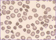
The patient in this case study was a male diagnosed with infantile nephrotic cystinosis at the age of eleven months. Nephrotic cystinosis is a rare autosomal recessive disorder resulting in the accumulation of cystine crystals primarily in the kidney as well as other organs, including the bone marrow. These crystals are almost never seen in the peripheral blood. Cystinosis presents between six and twelve months of age. The clinical characteristics include failure to thrive, progressive renal failure, loss of muscle function, photophobia and fair hair. Diagnosis is made by measuring the level of cystine in peripheral blood white cells.

By the age of eleven years, the patient’s condition had progressed to a stage which involved renal failure requiring peritoneal dialysis and frequent blood transfusions. At the age of thirteen, he received a renal transplant, the kidney being donated by a family member. He was then started on a regimen of immunosuppressive therapy.

By the age of twenty the patient was experiencing neurological abnormalities. He had difficulty in walking and swallowing, loss of memory and progressive loss of speech as well as diminished intellectual function. He continued to receive immunosuppressive therapy for his renal transplant.

At the age of twenty-six he presented to the Casualty Department. He had cervical and mediastinal lymphadenopathy and splenomegaly. A full blood count was performed with the following results:
Hb 115 g/L, WBC 5.4 x 10⁹/L and platelet 
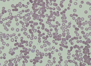
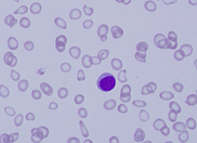
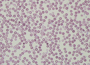
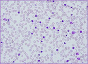
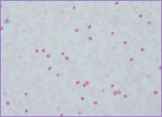
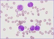
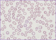
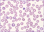
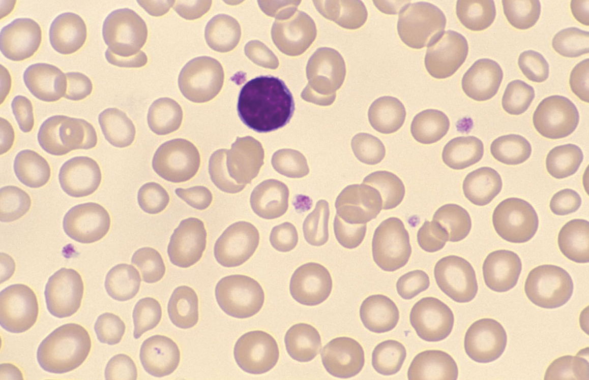
count 197 x 109/L. The peripheral blood 
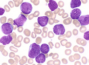
film revealed the presence of circulating blast 
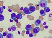
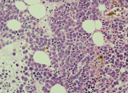
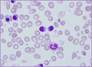
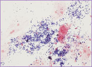
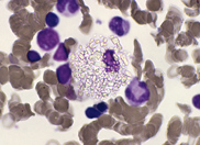
cells. A bone marrow biopsy was performed. The bone marrow was hypercellular with 70% blasts. The blasts had a high N/C ratio; the nuclei were variable from round to irregular and folded in shape. They contained one to four distinct nucleoli. Immunophenotyping was performed with the following results:

CD45+, HLA-DR, CD19+, cytoCD79a+, cytoCD3+, CD2+, CD5+, CD7+, CD38+ and CD117+.
CD4, CD8.

A diagnosis of T-cell lymphoblastic lymphoma/leukaemia was made.
CD2, 5 and 7 are the most commonly expressed markers in T-cell lymphoma/leukaemia. CD3 is often present in the cytoplasm but not on the surface. CD4 and CD8 may be expressed or may be negative.

Co-expression of CD79a and CD117 has been described on primitive T cell lineages however the expression of CD19, normally a B cell marker, is unusual.

A cytogenetic analysis on the available cells revealed a 46, XY karyotype. Subsequent fluorescence in situ hybridisation (FISH) studies showed no evidence of BCR-ABL 1 gene rearrangements.

Post-Transplant Lymphoproliferative Disease

BM showing macrophage laden with crystals of cystine
BM showing a heavy infiltration of lymphoblasts

Post-Transplant Lymphoproliferative Disorders (PTLD) are a well-known complication of immunosuppressive treatment after solid organ transplantation. Any lymphoma/leukaemia that occurs in the post-transplant population is considered to be a type of PTLD. The median time from transplant to diagnosis is fifteen years. The majority of post transplant lymphoma/leukaemias are B cell in origin and are associated with EBV infection. T-cell variants are very rare. The patient in this study was EBV positive.

The risk factors of PTLD are intensity of immunosuppression, type of organ transplanted, Epstein-Barr virus (EBV) seronegativity and the duration of the immunosuppression. EBV seronegativity pre-transplant is a powerful predisposing factor for the development of PTLD. Studies show that EBV seronegative patients experience a 10-76 fold greater incidence of PTLD when compared to seropositive counterparts.

The patient in this study died suddenly, two months post chemotherapy induction.

⬆ Back to Top

A case of haemolytic anaemia occurring in a young child

Case Study 22

A three-year-old female child presented at the casualty department. She had been unwell with tonsillitis for the past week. A full blood count revealed the following results:
Hb 67 g/L, WBC 24.2 x 10⁹/L and platelets 452 x 109/L.

The blood film was leucoerythroblastic with a reticulocytosis and a moderate number of spherocytes. There was an absolute neutrophilia of 14.0 x 10⁹/L. A preliminary diagnosis of autoimmune haemolysis was made. A direct antiglobulin test (DAT) was performed with a positive result. The LDH was raised at 666 IU/L demonstrating acute haemolysis. Urinalysis revealed dark brown urine with a small number of red cells.

As this was an acute presentation (the child was well two weeks previously) the possibility of paroxysmal cold haemoglobinuria (PCH) was considered. A Donath-Landsteiner test was performed and the result was positive.

PCH is a disease characterised by the sudden presence of haemoglobin in the urine after exposure to cold temperatures. It occurs mostly in young children under the age of five years. There is a male to female ratio of 2:1 to 5:1.

In 1904 Julius Donath and Karl Landsteiner described an autoimmune haemolytic anaemia characterised by the presence of a biphasic antibody that attaches to red cells in the cold inducing haemolysis when the red cells are subsequently warmed. The Donath- Landsteiner (D-L) antibody is a polyclonal IgG binding to various red cell antigens such as I, i, p and P on the red cell surface. The P antigen is its primary target. This polyclonal IgG anti-P autoantibody binds to red blood cell surface antigens in the cold. When the blood returns to the warmer central circulation, the red cells are lysed with complement, causing intravascular haemolysis. The attachment of C3d on the red cells results in a positive DAT. The resultant blood picture is that of a normochromic normocytic anaemia with spherocytes, reticulocytes, especially in the recovery phase, auto agglutination and nucleated red cells. Monocytes and granulocytes sometimes show erythrophagocytosis.

Peripheral blood (LP) showing auto agglutination
Peripheral blood (HP) showing spherocytes

The Donath-Landsteiner (D-L) antibody test is performed as follows:
The method involves preparing three tubes: 1) the patient’s serum, 2) a mixture of patient’s serum and normal serum and 3) normal serum. To each tube is added P-positive red cells at 4℃. After 30 minutes the tubes are then heated to 37℃ and observed visually for haemolysis, indicative of a positive result.

PCH is associated with upper respiratory and gastrointestinal symptoms. It may occur following measles, mumps, influenza, varicella zoster virus, cytomegalovirus, Epstein-Barr virus, adenovirus, parvovirus B19, Coxsackie A9, Haemophilus influenzae, Mycoplasma pneumoniae and Klebsiella pneumoniae. It also may occur post vaccination for measles.

PCH is a self-limiting disease. If promptly diagnosed and appropriately treated with supportive care, most patients recover within days to a few weeks. Supportive treatment may include pre-warmed packed red blood cells as well as steroids although steroids have not been shown to shorten the course of the disease.

The patient in this case study was transfused warm packed red blood cells. She remains well.

⬆ Back to Top

A case of plasma cell myeloma in a middle aged male

Case Study 23

A 53-year-old male was referred to the Haematology clinic with severe back pain together with a normochromic, normocytic anaemia for investigation. A full blood count was performed with the following results:
Hb 108 x g/L, WBC 4.5 x 10⁹/L, platelet count 227 x 109/L and ESR of 125 mm/hr.

The blood film confirmed a normochromic, normocytic anaemia with a marked increase in rouleaux formation. The white cells and platelets were normal in number and morphology.

Serum proteins were performed. A paraprotein IgGk of 91g/L was identified by immunofixation in the beta globulin region.

Bence Jones protein, 3.49 g/24hr, was found in the urine.

A bone marrow aspiration was performed. There was a heavy infiltrate of plasma cells displacing much of the normal haematopoietic tissue of the marrow. The plasma cells were large with eccentrically placed nuclei and basophilic cytoplasm. Binucleate forms were frequent. Mitotic figures were also increased.

A bone marrow trephine showed a similar picture.

A diagnosis of plasma cell myeloma was made.

Immunophenotyping on the marrow showed the following:
CD19, CD20, CD138+, CD38+, and CD79a+

Bone marrow trephine showing sheets of plasma cells

Bone marrow biopsy showing sheets of plasma cells
Bone marrow trephine showing a heavy infiltrate of plasma cells

Plasma cell myeloma is a neoplasm occurring more commonly in males. The median age at presentation is 50 years. The initial presentation is associated with bone pain, back, neck and pelvic pain. Radiographic findings reveal lytic lesions and osteoporosis. Pathological fractures can occur in up 70% of patients.

Plasma cell myeloma is diagnosed from the bone marrow. It is invariably associated with a paraprotein or M-protein in the serum and/or the urine. The protein in the urine is referred to as Bence Jones protein.

Symptomatic plasma cell myeloma is associated with CRAB: hypercalcaemia, renal insufficiency, anaemia and bone lesions.

The peripheral blood shows an increase in rouleaux formation. Plasma cells are found in approximately 15% of cases and only in small numbers.

The bone marrow shows an increase in plasma cells, more than 3% and sometimes up to 90%. Mature plasma cells are oval to round in shape with round eccentric nuclei. The cytoplasm is basophilic with a perinuclear hof. Crystals as well as globules of immunoglobulin are sometimes seen in the cytoplasm. Plasma cells appear in clusters or sheets within the marrow with preservation of normal haematopoiesis.

When the number of plasma cells in the peripheral blood exceeds 2 x 10⁹L the neoplasm is referred to as plasma cell leukaemia.

Plasma cell myeloma is usually incurable. It has a median survival of 3-4 years. Transplantation may be attempted to increase and prolong quality of life.

The patient in this case study remains well.

⬆ Back to Top

A Medical Dilemma

Case Study 24

An 8 year old child with Pfeiffer syndrome presented at the Paediatric Casualty Department. She had a two week history of severe pallor and progressive lethargy.

Pfeiffer syndrome is a genetic disorder characterised by craniosynostosis, visual impairment and ocular proptosis. It is thought to be associated with deficiencies associated with fibroblast growth factor receptors on chromosome 8 and 10.

This young child was of short statue. She had dysmorphic features and was blind. She demonstrated reduced exercise tolerance, unable to keep up with her siblings. She had a history of complex neurosurgical and plastic surgery. She had difficulty in eating solids hence her dietary intake was pureed solids; meats, fish and dairy products.

Her parents had become Jehovah Witnesses in 2006.

A full blood count was performed with the following results:
Hb 23 g/L, WBC 6.8 x 10⁹/L and Platelet count 259 x 109/L.
The RBC was 1.97 x10¹²/L, Hct 0.100, MCV 51.8 fL and RDW-CV 23.1%.
The Reticulocyte count was 1.5% and absolute count 29.4 x 10⁹/L.
The blood film revealed a marked microcytic, hypochromic anaemia with elliptocytes and pencil cells consistent with a severe iron deficiency anaemia.

Iron studies were performed with the following results:
Iron 1.2 µmol/L, Transferrin 3.8 g/L, Iron saturation 1.3% and Ferritin 1 µg/L.
Parvovirus B19 was negative and the Hb EPG was normal.

Bone marrow trephine showing sheets of plasma cells

Initial blood film with Hb 23 g/L
Post
Post transfusion, dimorphic blood film

The Medical Dilemma was now clearly an acute life threatening event. The child had cardiac signs and was at risk of cardiac failure or arrest. She was in need of intensive care. There was also a need to acknowledge the religious beliefs of the family. At this point a hospital administrator was called. The parents reluctantly consented for a blood transfusion. Meanwhile the child’s father contacted support from the Jehovah Witness community to attend the hospital. Consent to transfuse was withdrawn.

The possibility of an iron transfusion and administration of erythropoietin was discussed. However the Consultant Haematologist caring for the child was adamant that she required an urgent blood transfusion. At this point in time the hospital Chief Executive Officer was contacted as was a social worker, Department of Community Services and the Child Protection Consultant. The Consultant Haematologist was requested to attend the Supreme Court under section 174 of the Children and Young Persons Act 1998. However, provided the Consultant Haematologist was prepared to sign an affidavit that the child required a transfusion a visit to the court could be waived. An affidavit was signed and the child was transfused.

The post transfusion Hb was 125 g/l.

The Department of Community Services was not required in this case and the child was discharged.

⬆ Back to Top

A potentially very unwell full term neonate

Case Study 25

Late one evening, a full blood count was received on a full term neonate who had just been delivered by caesarean section. The clinical notes read ‘pallor’. The results from the analyser were as follows:
Hb 64 g/L, MCV 118.9 fL, reticulocyte count 13.4 % (254 x 109/L), WBC 16.2 x 10⁹/L and platelets 278 x 109/L.

A Hb result of 64 g/L is abnormally low for a newborn neonate; the normal range being 121 – 191 g/L in the above laboratory. A repeat specimen was requested. The Hb was now 60 g/L, confirming the initial result. Whilst the blood film was being stained a direct antiglobulin test (DAT) was performed with a negative result. Both the infant and mother were group O positive and an antibody screen performed on the mother was negative. The total bilirubin was 140 umol/L, NR (0-150 umol/L).

The infant was transfused with two units of packed cells.

Blood film (x 400)
Blood film (x 400)
Blood film (X1000)

The blood film revealed a nucleated red cell count (NRBC) of 674 NRBCs / 100 WBCs. The normal range being 1-25 NRBCs / 100 WBCs. The blood film image (x 400) demonstrates the markedly increased number of red cell precursors while the blood film images (x 1000) demonstrate marked dyserythropoiesis. Note the nuclear budding and cytoplasmic bridging, classical features of dysplasia.

A differential diagnosis on this infant would include a fetomaternal bleed or less probably Congenital Dyserythropoietic Anaemia (CDA). A Kleihauer test was performed on the mother to check for a fetomaternal bleed. The Kleihauer test was positive; the presence of 3.84 % fetal cells translated to a transplacental haemorrhage of 95.84 mls. Hence the initial diagnosis of a fetomaternal bleed was confirmed.

Kleihauer-Betke (X 400)

Some degree of fetomaternal transfusion occurs in approximately 50% of all pregnancies. The most frequently observed causes of occult haemorrhage prior to birth include abdominal or multiple trauma, amniocentesis in the third trimester, post external cephalic version, placental tumours and spontaneous haemorrhages. Haemoglobin values as low as 30 to 60 g/L have been recorded in infants who were born alive and survived.

The clinical manifestations of a fetomaternal haemorrhage depend on the rapidity with which it has occurred. If the haemorrhage has been prolonged, giving the fetus the opportunity to compensate for the anaemia, the infant may manifest only pallor at birth. However, if the haemorrhage is acute, the infant may be pale and sluggish, have gasping respirations and manifest signs of circulatory shock.

The blood film may also be informative. In an acute haemorrhage, the red cells are normochromic normocytic whereas in a more prolonged fetomaternal haemorrhage the red cells may be microcytic and hypochromic. Note that the NRBCs in this case exhibited ragged cytoplasm, a feature seen in iron deficiency. The MCV on this neonate was slightly raised at 118.9 fL, NR (101-117 fL). This was secondary to the significantly raised reticulocyte count of 13.4 % NR (4-7 %). In anaemia secondary to a fetomaternal haemorrhage the DAT test is negative and the infants are not jaundiced. The presence of a very high NRBC count is an index of fetal stress.

The diagnosis of a fetomaternal haemorrhage can be made with certainty by demonstrating the presence of fetal cells in the maternal circulation. This is done by performing the Kleihauer-Betke test. The Kleihauer image above demonstrates the presence of acid-resistant fetal red cells on a background of acid sensitive adult red cells that appear as ghost cells. Note that the Kleihauer-Betke method has been replaced by a flow cytometry test for the estimation of HbF in many laboratories.

The infant in this update maintained a stable haemoglobin post transfusion hence a potential diagnosis of CDA was not pursued. Mother and infant went home several days after confinement.

⬆ Back to Top

Failure to thrive

Case Study 26

A seven weeks old infant was admitted to hospital with a history of ‘failure to thrive’. A full blood count was performed with the following results:
Hb 123 g/L, WBC 10.2 x 10⁹/L Platelets 586 x 109/L

Examination of the blood film revealed a marked number of acanthocytes. A provisional diagnosis of abetalipoproteinaemia was made.

The following chemistry tests were performed:

Cholesterol 1.4 mmol/L NR (3.0-5.5)
Triglyceride <0.1 mmol/L NR (<2.0)
Apolipoprotein A1 0.78 g/L NR (1.10-2.00)
Apolipoprotein B 0.07 g/L NR (0.60-1.25)
Vitamin A 0.4 umol/L NR (0.5-1.8)
Vitamin E <1 umol/L NR (5-14)
Faecal fat 38 mmol/day NR (0-3.4)

The above results confirmed a diagnosis of abetalipoproteinaemia.

Peripheral blood film showing a marked number of acanthocytes

Abetalipoproteinaemia is a rare autosomal recessive disorder characterised by the presence of acanthocytic red cells in the peripheral blood. The primary defect is due to a mutation and lack of activity in the microsomal triglyceride transfer protein needed to bind lipids to the ?-apolipoprotein in plasma. Intestinal absorption of lipids is defective. The plasma levels of cholesterol and triglyceride are extremely low and ?-apolipoprotein undetectable. There is an increase in sphingomyelin in the outer half of the red cell membrane bilayer increasing the surface layer of the cell. This ?-apolipoprotein defect leads to the production of acanthocytic red cells, about 50-90% of the red cells being acanthocytes. The sphingomyelin accumulates with cell ageing thus the nucleated precursor red cells and reticulocytes are not affected while the older red cells are affected.

As a result of fat malabsorption and the absence of low density lipoproteins, which transport vitamin E, the red cells of patients with abetalipoproteinaemia are markedly deficient in vitamin E. The level of vitamin A is also reduced.

Abetalipoproteinaemia usually presents in early childhood with diarrhoea and failure to thrive and is characterised clinically by fat malabsorption, spinocerebellar degeneration, pigmented retinopathy and acanthocytosis. The initial neurologic manifestations are loss of deep-tendon reflexes, ataxia and development of a spastic gait. They also develop retinitis pigmentosa which ultimately progresses to near-blindness. The neurological abnormalities present between five and ten years of age and continue until death in the second or third decade. Despite the marked acanthocytosis and vitamin E deficiency seen in these patients, anaemia and haemolysis are mild. The haemoglobin levels are invariably normal.

Treatment consists of a low-fat, high caloric, vitamin-enriched diet accompanied by large supplemental doses of vitamin E. It is imperative that treatment be initiated as soon as possible to delay the onset of the neurological symptoms. New therapies for this debilitating disease are needed.

⬆ Back to Top

Acute promyelocytic leukaemia at 28 weeks gestation

Case Study 27

A 29 year old female presented to the Casualty Department feeling generally unwell. At 28 weeks gestation she was hypertensive with proteinuria. She had with her an Advanced Health Care Directive stating that no transfusions of whole blood, red cells, white cells, platelets or plasma be administered to her. She was admitted with a presumptive diagnosis of pre-eclampsia.

A full blood count and coagulation studies were performed with the following results:

Hb 103 g/L WBC 10.5 x 10⁹/L Platelets 24 x 109/L
PT 15.8 sec INR 1.2 APPT 27.4 sec
Fibrinogen 1.4 g/L D- DIMER LIA >20.00 ug/ml

She was severely thrombocytopenic with disseminated intravascular coagulation (DIC). The blood film and differential count showed the presence of 15% blast cells and 55% abnormal promyelocytes. A diagnosis of acute promyelocytic leukaemia (APL) was made. A bone marrow aspiration was performed. The differential count on the bone marrow showed the presence of 86% abnormal promyelocytes. Cytogenetics showed a t(15;17). FISH confirmed a PML/RARA fusion. Immunophenotyping was as follows:
CD45+, HLA-DR-, CD11b-, CD13+, CD15-, CD33+, CD34-, CD64+, CD117+, CD2+, CD22+ and MPO+

The above patient was transferred to a ward under joint obstetric and haematology care. A number of issues were before this medical team. They were presented with a patient with APL, DIC and severe thrombocytopenia. She was 28 weeks pregnant with pre-eclampsia and from deeply-held conviction, refused blood product support. Therefore performing a caesarean section was not safe. She was given steroids for fetal lung maturity as well as all trans retinoic acid (ATRA), the drug of choice for the treatment of APL.

Over the next few days the white cell count steadily rose as did the urea, creatinine and liver function tests. Her hypertension was difficult to control. Labour was induced with failure to progress. On the ninth day after admission, a stillborn fetus was delivered vaginally. There was minimal blood loss. It is thought that the stillborn infant was directly related to the acute renal failure in the mother.

Peripheral blood film collected on day ten showing the presence of abnormal, bilobed promyelocytes with granular cytoplasm.
Peripheral blood film collected on day ten showing the presence of abnormal, bilobed promyelocytes with granular cytoplasm. The white cell count was 61.0 x 10⁹/L

By day ten the patient started to deteriorate. The white cell count had increased to 61 x 109/L, the Hb was 48 g/L and platelet count 17 x 109/L. Her renal function had deteriorated. The blood urea was 24.0 mmol/L and creatinine 282 mmol/L. The liver function became acutely deranged – GGT 59 U/L, AST 80 U/L and ALT 369 U/L.

She was admitted to intensive care where she was intubated. Her neurological function deteriorated. She was now hemiplegic. By day twelve there was further deterioration in her neurological function. On day thirteen treatment was withdrawn.

There are two types of APL. The hypergranular or typical type characterised by abnormal promyelocytes whose cytoplasm is densely packed with granules and Auer rods and the hypogranular or microgranular type whose cytoplasm shows a paucity or absence of granules and occasional Auer rods. The granules in these abnormal promyelocytes contain procoagulant mediators which, when released into the circulation, cause a life-threatening coagulopathy or DIC. The dilemma with the above patient was her severe thrombocytopenia, DIC and her refusal to accept blood products. Current therapy of ATRA combined with chemotherapy results in 70 to 80% patient survival and disease-free outcome after five years.

The initial visit by this patient to the hospital was to surrender an Advanced Health Care Directive stating that she would not accept a transfusion of red cells, white cells or platelets. As an adult she is able to make this decision. In the case of a child, a court order would ensure that the child be treated appropriately.

⬆ Back to Top

At last a Haematology Update with a happy ending!

Case Study 28

In February 1999 a 29 year old female attended the Haematology Clinic at the Prince of Wales Hospital. She had a history of anaemia, neutropenia and thrombocytopenia. She was passing dark urine. A full blood count, reticulocyte count and coagulation studies were performed.
The results were as follows:

Hb 66 g/L Retic count 16.20% (absolute count 356.40 x 10⁹/L)
MCV 113.6 fL WBC 3.1 x 10⁹/L Platelets 55 x 109/L
PT 41.5 sec INR 4.2 APTT 45.7 sec
Fibrinogen 5.6 g/L D-DIMER LIA <0.19 mg/L

A urinary haemosiderin was performed which was strongly positive.

A clinical chemistry profile was also performed:
The LDH was raised at 3436 U/L.

The test results, together with the clinical symptoms of the patient indicated that she had a severe haemolytic anaemia suggestive of Paroxysmal Nocturnal Haemoglobinuria (PNH). A Ham’s test was performed. This is an acidified serum test which detects a population of red cells with sensitivity to complement-mediated lysis. The Ham’s test was positive. A diagnosis of PNH was confirmed on the above patient.

PNH is a rare, acquired, clonal disorder of the bone marrow characterised by a total or partial lack of proteins normally attached to the haemopoietic cell membrane by the glycosylphosphatidylinositol (GPI) anchor. This defect is due to a somatic mutation of the phosphatidylinositol glycan class A (PIG-A) gene on the X chromosome which encodes for a protein needed for the synthesis of GPI anchor. Without GPI anchor, an essential group of membrane proteins are either reduced or absent in all haemopoietic cell lines. These proteins include complement-regulating surface proteins namely decay-accelerating factor (DAF) or CD55, membrane inhibitor of reactive lysis protein (MIRL) or CD59 and the homologous restriction factor (HRF) or CD8 binding protein. All of these proteins interact with complement, particularly C3b and C4b, protecting cells against lysis. In their absence, haemopoietic cells are exposed to complement leading to uncontrolled amplification of the complement cascade and destruction of the red cell membrane leading to intravascular haemolysis.

Peripheral blood film showing a raised MCV secondary to an increased reticulocyte response
Figure 1a: Peripheral blood film showing a raised MCV secondary to an increased reticulocyte response ( x 1000)
Strongly positive urinary haemosiderin
Figure 1b: Strongly positive urinary haemosiderin ( x 400)

PNH cells are classified as PNH Type I, Type II and Type III cells. PNH Type I cells have normal levels of CD59; PNH Type II cells have reduced levels while PNH Type III cells completely lack CD59 expression.

Patients with PNH present with symptoms of haemolysis, namely, low haemoglobin, raised reticulocyte count and raised lactate dehydrogenase (LDH). The DAT is negative as there are no antibodies involved. They also present with isolated or multiple cytopenias and/or a history of thrombosis.

Note that in 1999 the Ham’s test was used to make the diagnosis of PNH. Today flow cytometry must be used to make that diagnosis. The presence and size of both red blood cell and granulocyte clones as defined by reduced or absent GPI-linked proteins, must be determined. The Australian Flow Cytometry Group has issued guidelines for the most appropriate flow testing (AFCG Guidelines at www.afcg.org.au). The guidelines recommend that CD59 expression should be determined on red blood cells and that for granulocytes and monocytes the first step is a lineage gate (eg CD33/SS) followed by expression of CD24/FLAER (granulocytes) and CD14/FLAER (monocytes).

Until recently, the treatment for PNH was supportive. Patients were managed with iron and folate supplementation. They were also transfused when appropriate. Transfused red cells do not lack GPI anchor thus are not vulnerable to complement lysis. About 50% of patients responded to corticosteroids. Allogeneic stem cell or bone marrow transplantation was and still is the only cure for PNH.

In March 2007 the use of eculizumab (Soliris) was approved by the FDA. Eculizumab is a monoclonal antibody that targets and prevents cleavage of the terminal complement protein C5. In so doing it inhibits other complement components thus preventing haemolysis. Eculizumab has given patients with PNH a significant improvement in quality of life as well as a marked reduction in the frequency of thrombotic episodes. Many patients are no longer transfusion dependent. Long term treatment with eculizumab is well tolerated however it comes with significant cost consideration.

The above patient was commenced on eculizumab in 2009. It is now September 2010 and she is 39 years old. She has had a remarkable improvement in her quality of life whilst on eculizumab, so much so that she delivered a healthy baby girl on the 23rd of September. At last a Haematology Update with a happy ending!
Figure 2.
A. CD59 expression on red blood cells of the patient in 2010 showing a Type III PNH clone of 56.5%, a small Type II clone (8.3%) and a clone of Type I red
B. A PNH monocyte clone with reduced CD14 and FLAER expression – about 95% of monocytes.
C. A PNH granulocyte clone with reduced CD24 and FLAER expression – about 95% of granulocytes.

Figure 2

⬆ Back to Top

Severe anaemia in a 22 year old female

Case Study 29

A 22 year old female, found to have severe anaemia by her General Practitioner, presented at the Haematology Clinic for further investigative studies. A full blood count and haemoglobin electrophoresis (EPG) were performed .

The results were as follows:

Hb 76 g/L 115-165
Hct 0.244 0.37-0.47
MCV 83.3 fL 80-100
MCH 25.9 pg 26.5-33.0

The blood film was mildly microcytic and hypochromic. There was an occasional nucleated red cell present while many of the red cells showed coarse basophilic stippling.
The EPG results were as follows:
Hb EPG Cellulose Acetate (pH 8.6) No abnormal band detected.

Hb A2: 2.6 % 2.0-3.5 %
Hb F: <1.0 % <1 %

Hb H inclusions: not detected
No abnormality detected.
Iron studies were normal in this case.
A bone marrow aspirate was performed. The marrow was hypercellular with increased erythropoiesis and mild dyserythropoiesis. No abnormal sideroblasts were seen.

As coarse basophilic stippling in the red cells is a feature of lead poisoning, a lead level was performed. The lead was found to be 5.0 umol/L with a normal range of 0-0.5 umol/L. The level of mercury was also examined and found to be <1 nmol/L with a normal range of ≤50 nmol/L.

Blood film showing coarse basophilic stippling

After questioning the patient in this case study it became evident that the patient had been taking a medication supplied to her by her Indian doctor. The medicine was in a powder form. A sample of this medication was sent for analysis which showed that the powder contained 12 % lead.
Patients with lead poisoning clinically present with abdominal colic and constipation, peripheral neuropathy and anaemia. The anaemia is invariably a hypochromic microcytic anaemia as the ingestion of lead interferes with haem synthesis. It does so by inhibiting several enzymes directly involved with haem synthesis. Pyrimidine 5′-nucleotidase is one such enzyme.

In its absence, pyrimidine nucleotides accumulate in the red cells, preventing iron from being incorporated into haem at a normal rate. This leads to a shortened red cell life span resulting in a mild haemolytic anaemia. The characteristic feature on the blood film in lead poisoning is coarse basophilic stippling in the red cells. The hypochromic microcytosis is not necessarily associated with iron deficiency.

The patient in this case study was treated with Meso-2-3-dimercaptosuccinic acid (DMSA) commonly known as succimer, a compound approved by the FDA in the 1960’s for the chelation or removal of heavy metals, particularly lead and mercury. She was initially given a three week course of succimer, 400 mg in the morning and 600 mg in the evening. Her haemoglobin began to rise while the level of lead began to fall. Two months later a similar course of succimer was administered. By the end of the second course the lead level was 0.8 umol/L, still significantly raised. The haemoglobin had returned to normal.

The Public Health Act (1991) states that a lead level of ≥0.72 umol/L must be notified and that repeat testing should occur after three months to assess the effectiveness of treatment.

⬆ Back to Top

A case of necrotising fasciitis in a 56 year old male

Case Study 30

A 56 year old male was transferred from a regional hospital to the intensive care ward at the Prince of Wales Hospital. He had extensive necrosis extending from the scrotum to his flank, axilla, and shoulder and spreading along his arm. He had necrotising fasciitis and was to have surgical debridement. He was too unwell for the Hyperbaric Chamber.

A full blood count was received in the laboratory. The results were as follows:

Hb 89 RR (130-180) g/L
Hct 0.262 RR (0.40-0.54)
MCV 87.9 RR (80-100) fL
MCH 29.9 RR (26.5-33.0) pg
WBC 39.9 RR (3.5-11.0) x 10 9/L
Plats 80 RR (150-400) x 10 9/L

The blood film showed an absolute neutrophilia with marked toxic granulation; moderate numbers of microspherocytes and thrombocytopenia. These features are classically found in Clostridium perfringens infection. This was a case of severe septicaemia and haemolysis secondary to necrotising fasciitis. See figure 1.

The term necrotising fasciitis describes a condition of rapidly spreading infection, usually located in fascial planes of connective tissue resulting in tissue necrosis. Fascial planes are bands of connective tissue that surround muscles, nerves and blood vessels. The speed with which necrotising fasciitis spreads is directly proportional to the thickness of the subcutaneous layer.

Many types of bacteria can cause necrotising fasciitis ( e.g., Group A streptococcus (Streptococcus pyogenes), Staphylococcus aureus, Clostridium perfringens, Bacteroides fragilis, Aeromonas hydrophila). The disease is classified as either Type I (polymicrobial) caused by a number of different organisms or Type II (monomicrobial) caused by a single organism. The causative organism may be aerobic or anaerobic.
The frequency of necrotizing fasciitis has been on the rise due to an increase in immunocompromised patients with diabetes mellitus, cancer, alcoholism, vascular insufficiencies, organ transplants, HIV infection and also occurs in patients with neutropenia
The mean age of a patient with necrotizing fasciitis is 38-44 years. The disease rarely occurs in children. Paediatric cases have been reported from resource-poor nations where poor hygiene is prevalent.

The patient in this case study was diagnosed with Clostridium perfringens induced necrotising fasciitis. Clostridium perfringens is a saprophytic organism inhabiting the bowel and genital tract. It has no pathological significance in the absence of clinical infection. Clostridium perfringens produces at least 12 antigenic protein toxins, the most common of which is the alpha toxin. These toxins react with lipoprotein complexes on cell surfaces, liberating potent haemolytic substances known as lysolecithins which result in cell lysis, hence the presence of microspherocytes on the blood film. This process leads to a severe haemolytic anaemia. Acute renal and hepatic failure develops leading to death in as short a period as 12 hours if not treated immediately.

The Chemistry results on this case were as follows:

Urea 10.1 mmol/L RR (2.9-7.1)
Creatinine 152 umol/L RR (60-110)
Bilirubin total 47 umol/L RR (0-25)
ALP 106 U/L RR (38-126)
GGT 71 U/L RR (0-50)
AST 89 U/L RR (<45)
ALT 72 U/L RR (<45)

The basis of treatment is surgical debridement of necrotic tissue and antibiotic therapy. In severe infections, hyperbaric oxygen is an important adjunct in the treatment of necrotising fasciitis. Massive doses of benzyl penicillin are administered intravenously. Should the patient be allergic to penicillin, metronidazole is effective in high doses.

The patient in this case study died within 24 hours of having been admitted into the ICU ward.

⬆ Back to Top