Microscopic Haematology / Case Studies

A Practical Guide for the Laboratory

Transparent Logo

Differential diagnosis of hereditary stomatocytosis and Southeast Asian ovalocytosis

Case Study 1

The blood film of a patient with either hereditary stomatocytosis or Southeast Asian ovalocytosis will show stomatocytes as a common feature. However, there are important differences in the red cell parameters that permit one to differentiate morphologically between the two disorders.

Hereditary Stomatocytosis
Hereditary stomatocytosis
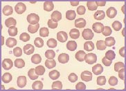
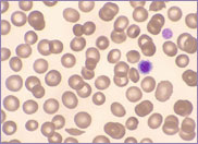
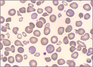
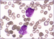
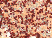
Macrocytic stomatocytes as well as post-splenectomy changes, in a case of hereditary stomatocytosis (hydrocytosis).
Southeast Asian ovalocytosis
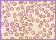
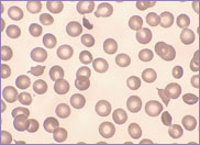
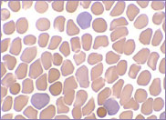
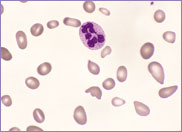
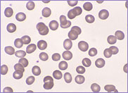
Oval-shaped stomatocytes, some with two transverse slits, in a case of Southeast Asian ovalocytosis.

Hereditary stomatocytosis, often referred to as hereditary hydrocytosis, is a very rare autosomal dominant disorder, characterized by a defect in the Na+ / K+ exchange pump. There is an imbalance between the amount of Na+ entering the red cell and the amount of K+ exiting the cell. The red cell swells as it gains cations and water, transforming from a discocyte to a stomatocyte. These swollen cells are osmotically fragile. They become rigid, requiring extremely large amounts of energy to protect them against lysis.

The red cells of hereditary stomatocytosis lack stomatin (protein 7.2b), an integral red cell membrane protein. The exact function of stomatin is unknown, but it is clearly important in regulating membrane sodium permeability.

The laboratory findings in hereditary stomatocytosis include a macrocytic anaemia. The blood film shows stomatocytes and round macrocytes. The mean cell volume (MCV) is characteristically elevated in the range of 110-150 fL, and there is a correspondingly low mean corpuscular haemoglobin concentration (MCHC) in the range of 240-300 g/L. An elevated red cell Na+ concentration and a reduced K+ concentration, as well as an increased total monovalent cation content, are noted. These abnormal red cells are sequestrated on passing through the spleen, giving rise to extravascular haemolysis. The haemolytic anaemia is severe with a reticulocytosis of >10%. Splenectomy has proven to be the most beneficial treatment in reducing the degree of haemolysis.

Southeast Asian ovalocytosis is also an autosomal dominant disorder. It is very prevalent, occurring in up to 30% of aboriginal people from New Guinea, Malaysia, and Melanesia.

The red cells of Southeast Asian ovalocytosis have a characteristic appearance on the blood film. They are often described as stomatocytic elliptocytes. Instead of being discocytes, they have a slit-like area of central pallor. A small proportion of these stomatocytes have two transverse slits, giving the appearance of double stomatocytes. The red cell membrane of these stomatocytes is very rigid. This abnormality results from increased ankyrin binding and decreased protein 3 mobility, leading to the production of rigid red cells. This rigidity is a protective mechanism against all strains of malaria. There is no abnormality in the red cell Na+/K+ exchange pump.

The laboratory features of Southeast Asian ovalocytosis are unremarkable. The stomatocytes have a normal lifespan; hence, there is no anaemia. The MCV and MCHC are both characteristically within the normal range. In fact, nowadays, with the use of cell analyzers to auto-validate results without examining blood films in the presence of normal blood cell parameters, laboratories diagnose fewer cases of this asymptomatic condition.

⬆ Back to Top

Help for HELLP

Case Study 2

A 30-year-old pregnant female presented with severe preeclampsia at 35 weeks gestation. A full blood count and clinical chemistry profile were performed, and the following results were noted:

  • Hb 123 g/L, WBC 17.7 x 10⁹/L, platelet count 99 x 109/L, and D-Dimer 9.6 µg/ml.
  • Total bilirubin 60 µmol/L, ALT 839 U/L, AST 1739 U/L, and LDH 2792 IU/L.

Four hours later, the above tests were repeated with the following results:

  • Hb 117 g/L, WBC 15.4 x 10⁹/L, platelet count 55 x 109/L, and D-Dimer 10.4 µg/ml.
  • Total bilirubin 98 µmol/L, ALT 791 U/L, AST 2452 U/L, and LDH 3500 IU/L.
  • The initial blood film showed the presence of an occasional schistocyte or sharp fragmented red cell. The presence of so few schistocytes, together with a rapidly falling platelet count, was suggestive of a microangiopathic process. The blood film prepared 4 hours later showed an increase in the number of schistocytes. A diagnosis of HELLP was made for this patient.
Occasional schistocyte and large platelets present on film
Initial presentation of HELLP
Occasional schistocyte and large platelets present on film
Slight increase in the number of schistocytes and reduction in platelets present on film
HELLP 4 hours post diagnosis
Slight increase in the number of schistocytes and reduction in platelets present on film

The HELLP syndrome (haemolysis, elevated liver enzymes, and low platelet count) is a multisystem disorder occurring in severe preeclampsia-eclampsia and is characterized by a microangiopathic haemolytic anaemia, hepatic dysfunction, and thrombocytopenia. In many instances, this progresses to disseminated intravascular coagulation (DIC).

The HELLP syndrome affects an estimated 4% to 12% of patients with severe preeclampsia. Its presence is recognized by the onset of symptoms and signs of hepatic involvement such as epigastric pain, nausea, and malaise, and on examination, right upper-quadrant tenderness. Because the HELLP syndrome is insidious in its onset, the early, milder form may be missed unless appropriate laboratory assessments—namely, FBC, LFTs, urea and creatinine, LDH, and DIC screen—are performed. Thus, the HELLP syndrome is often advanced before an accurate diagnosis is made.

The HELLP syndrome affects primiparous and multiparous women in the third trimester of pregnancy. Delivery of the fetus is the appropriate initial treatment. The syndrome remains active after delivery and appears to reach maximal intensity during the 24-48 hour post-delivery period when thrombocytopenia, ALT, AST, and LDH all reach their peak. The pathogenesis is not clear. As a result of fetomaternal cell traffic, fetal products induce the release of agonists in the mother, causing vascular endothelial damage and platelet activation. This induces DIC, leading to fibrin deposition and obstruction to blood flow. Interference with flow in the liver leads to the high liver enzyme levels observed in the HELLP syndrome. Red cells are damaged as they pass through fibrin-coated vessels, resulting in haemolysis. Thrombocytopenia is due to both platelet consumption and destruction. There is a correlation between the platelet count and the severity of the HELLP syndrome. Platelet counts below 50 x 109/L are associated with greater maternal morbidity.

The blood picture in the HELLP syndrome is characterised by the presence of schistocytes together with thrombocytopenia. The summation at the end of the blood film report should always read 'blood film consistent with a microangiopathic process'.

Plasmapheresis improves the HELLP parameters in non-responsive cases in the postpartum period. It removes a large variety of biologically active substances.

⬆ Back to Top

Hairy Cell Leukaemia

Case Study 3

A 58 years old female presents with a history of fatigue, easy bruising and recurrent infection. She is found to have gross splenomegaly. A full blood count is performed and the following results noted:

Hb 143 g/L, WBC 45.0 x 10⁹/L and platelet count 91 x 109/L.

The peripheral blood film showed 84% abnormal lymphocytes with irregular cytoplasmic projections and 16% neutrophils. A diagnosis of the variant form of hairy cell leukaemia (HCL-V) is made.

Hairy cell leukaemia-variant Peripheral blood
Hairy cells showing round, centrally placed nuclei and blue-grey cytoplasm with fine projections
Hairy cell leukaemia-variant Bone marrow
Hairy cells with round nuclei with variable amounts of blue-grey cytoplasm and fine cytoplasmic projections

Hairy cell leukaemia is an indolent malignant lymphoproliferative disease of B-cell origin. It occurs more often in men than in women (ratio 4:1). The classical form presents with pancytopenia, including neutropenia, and a mean white cell count of less than 4 x 10/L. In 15%-20% of cases, there is a variant or leukaemic form with a mean white cell count of 88 x 10/L. The variant form is not neutropenic.

The hairy cells on the peripheral blood film in the classical form vary in size from 10-20 µm in diameter. The cell cytoplasm stains a pale blue-grey colour with many fine hair-like projections around the entire circumference of the cell. This is in contrast to the projections found on the cells of splenic marginal zone lymphoma which are thin and short and unevenly distributed, often concentrated towards one pole of the cell. In classical hairy cell leukaemia, the nucleus is often eccentric and is round to oval in shape. The hairy cells in the variant form are smaller with a diameter of 10-15 µm. The nuclei are generally placed centrally rather than eccentrically. In both forms, there may be few recognisable hairy cells in the peripheral blood and diagnosis relies on a bone marrow study. The marrow trephine shows a patchy or diffuse interstitial infiltrate characterized by a halo of pale-staining cytoplasm surrounding a sea of round to oval hairy cell nuclei.

Flow cytometry performed on hairy cell leukaemia shows expression of pan-B-cell antigens CD19, CD20 and CD22 but are negative for CD5 and CD23 (both being positive in B-CLL). Almost all cases of classical hairy cell leukaemia also express CD103, CD11c and CD25. In the variant form of hairy cell leukaemia, the cells are negative for CD25 (a pre B antigen) and positive/negative for CD103.

Cytochemistry can be used to demonstrate the hairy cells on a blood film. In 95% of cases of classical hairy cell leukaemia, the cells show strong tartrate-resistant acid phosphatase (TRAP) activity whereas in the variant form, only 60% show TRAP activity and it is only weak. Thus TRAP activity studies are not really useful to confirm a diagnosis of hairy cell leukaemia.

There are no specific cytogenetic changes in hairy cell leukaemia - add(14)(q32) occurs in 20% of cases, del(6)(q23) occurs in 10% of cases and del(14)(q22;q32) occurs in 10% of cases.

Patients with the variant form of hairy cell leukaemia have a median survival of 5 years. Those with the classical form have a longer survival. Some patients with mild disease do not require treatment and have a prolonged survival. Complications in hairy cell leukaemia include infection due to neutropenia and immune dysfunction, anaemia due to bone marrow infiltration and bleeding due to thrombocytopenia.

⬆ Back to Top

Hereditary Pyropoikilocytosis

Case Study 4

A 1 day old neonate presents with extreme jaundice at birth. A full blood count and chemistry profile are performed with the following results:

WBC 20.7 x 10⁹/L, Hb 114 g/L, MCV 86.0 fL, MCH 28.5 pg and platelet count 243 x 109/L.
Total bilirubin 403 µmol/L and conjugated bilirubin 283 µmol/L.

The blood film shows extreme poikilocytosis with micropoikilocytes, microspherocytes, triangular fragments, budding red cells and polychromasia. It is noted from the blood film, that the platelet count is falsely elevated and the MCV is very low for age. (The MCV range for a 1day old full term neonate is 101 – 117 fL). The extreme poikilocytosis is the major contributing factor to the very low MCV. A diagnosis of hereditary pyropoikilocytosis is made on the blood film.

A healthy 1 day old term neonate
Macrocytic red cells, polychromasia with reticulocyte count ranging from 3%-7%, occasional target cell, spherocyte and NRBC
Hereditary pyropoikilocytosis in a 1 day old term neonate
Marked numbers of poikilocytes and micropoikilocytes, microspherocytes, triangular fragments, budding red cells and polychromasia

Hereditary pyropoikilocytosis is an autosomal recessive disorder characterised by severe haemolytic anaemia with thermal instability of the red cells. It forms part of a group of haemolytic anaemias due to red cell membrane abnormalities. This group also includes hereditary elliptocytosis, Southeast Asian ovalocytosis and hereditary spherocytosis. Hereditary pyropoikilocytosis is found predominantly in Africans and African Americans but has also been seen in Caucasians and Arabs. The disease presents in the neonate with anaemia and jaundice and is diagnosed from the blood film that shows bizarre poikilocytes, micropoikilocytes, microspherocytes, triangular fragments and budding red cells. These red cell changes are caused by mechanical instability of the red cell skeleton due to defective binding of the spectrin chains. Erythrocyte spectrin maintains the cellular shape and provides structural support for the lipid bilayer of the red cell membrane. Disruption of the spectrin complex leads to disorders characterised by abnormally shaped red cells. Hereditary elliptocytosis is due to a gene mutation resulting in a qualitative spectrin abnormality. Hereditary pyropoikilocytosis co-inherits this qualitative spectrin abnormality as well as a second mutation that causes a quantitative spectrin deficiency. Thus hereditary elliptocytosis and hereditary pyropoikilocytosis are closely related disorders. Often one parent or a close relative of the child with hereditary pyropoikilocytosis will have hereditary elliptocytosis.

Thermal sensitivity and osmotic fragility tests may be performed to aid in the diagnosis of hereditary pyropoikilocytosis. In hereditary pyropoikilocytosis, the red cells demonstrate thermal autohaemolysis and increased osmotic fragility. The spectrin in normal red cells denatures at temperatures between 49ºC and 50ºC whereas in hereditary pyropoikilocytosis, the spectrin denatures at temperatures between 45ºC and 46ºC. Prolonged exposure at 37ºC will also induce fragmentation in hereditary pyropoikilocytosis.

The clinical course of hereditary pyropoikilocytosis is variable although the degree of haemolysis wanes with time. Splenectomy has been shown to improve the anaemia but has no effect on the abnormal red cell morphology.

⬆ Back to Top

Acute Promyelocytic Leukaemia

Case Study 5

A 50 year old female presents with lethargy, bruising and bleeding gums. A full blood count is performed and the following results noted:

  • Hb 74 g/L
  • WBC 2.3 x 10⁹/L
  • Platelet count 28 x 109/L

The coagulation results are:

  • PT 16.9 sec
  • INR 1.3
  • APTT 27.1 sec

The blood film is leucoerythroblastic with 48% promyelocytes. Many of the promyelocytes are abnormal with bilobed and reniform shaped nuclei. The cytoplasm is densely packed with large azurophilic granules as well as Auer rods and bundles of Auer rods (faggots).

A provisional diagnosis of acute promyelocytic leukaemia (APL) is made.

Acute promyelocytic leukaemia peripheral blood
Abnormal promyelocytes with hypergranulated cytoplasm and Auer rods
Acute promyelocytic leukaemia bone marrow
Abnormal promyelocytes with hypergranulated cytoplasm and some bilobed nuclei

Acute promyelocytic leukaemia may occur at any age although it predominantly occurs in adults. There are two types: the hypergranular or 'typical type' and the hypogranular or 'microgranular' type. Both are characterised by the presence of abnormal promyelocytes in the peripheral blood and bone marrow. The promyelocytes in the hypergranular type have nuclei, which vary greatly in both size and shape. They are often bilobed and reniform or kidney shaped. The cytoplasm is densely packed with large azurophilic granules, which may obscure the nucleus. Auer rods and bundles of Auer rods (faggots) are invariably present. The promyelocytes in the microgranular type have the same characteristic nuclear shape however the cytoplasm shows a paucity or absence of granules. This is due to the submicroscopic size of the azurophilic granules. Auer rods and bundles of Auer rods may also be seen in the cytoplasm.

The bone marrow in APL is hypercellular.

The promyelocytes of APL contain procoagulant material which, when released into the circulation, causes disseminated intravascular coagulation (DIC). Coagulation studies should always be performed when a case of APL is suspected from the blood film.

Flow cytometry performed on APL shows expression of the myeloid markers CD 33 (homogeneous and bright) and CD 13 (heterogeneous). HLA-DR and CD 34 are both negative, demonstrating the presence of a differentiating population of cells.

The myeloperoxidase (MPO) reaction is strongly positive in both the hypergranular and hypogranular type. The reaction product often masks the nucleus as well as the cytoplasm.

Acute promyelocytic leukaemia shows the characteristic t(15;17)(q22;q12). This translocation results from the breakage and reunion of bands 15q22 and 17q12. It is a balanced translocation, which is both specific and diagnostic for acute promyelocytic leukaemia. The standard 15;17 translocation has been analysed in considerable molecular detail. It has been shown that, as a result of this translocation, the truncated retinoic acid receptor alpha gene (RARα) on chromosome17q12 has moved to chromosome 15q22, where it has fused with a gene called PML, giving rise to a new hybrid gene called PML-RARα.

Acute promyelocytic leukaemia stands alone when it comes to treatment. It responds to all-trans-retinoic acid (ATRA) which induces remission by differentiating the leukaemic promyelocytes. This remarkable response to ATRA is directly related to the PML-RARα rearrangement in APL patients.

Treatment with ATRA is combined with cytotoxic chemotherapy.

The World Health Organization (WHO) describes three variant translocations associated with APL. They are t(11;17)(q23;q21), t(5;17)(q32;q12) and t(11;17)(q13;q21).

t(11;17)(q23;q21) is morphologically different from the other variant translocations in that the promyelocytes resemble the shape of normal promyelocytes. The nuclei are round. The cytoplasm is hypergranular and usually lacks Auer rods. There may be increased numbers of pelgeroid forms present. It also does not respond to treatment with ATRA.

⬆ Back to Top

Fever in a 12 month old child. Could it be sepsis or could it be Kawasaki disease?

Case Study 6

A 12 months old child presents with an acute febrile illness. A full blood count, ESR and blood cultures are performed and the following results noted:

Hb 90 g/l, WBC 18.6 x 10⁹/l, Platelet count 720 x 109/l and ESR 110 mm/hr.

The blood cultures show no growth after 7 days.

The blood film shows a normochromic, normocytic anaemia with marked rouleaux formation. The neutrophils show increased granulation and vacuolation. They also show cytoplasmic swelling, a characteristic feature of Kawasaki disease. A marked thrombocytosis is present.

Could the diagnosis be sepsis or could it be Kawasaki disease?

Acute promyelocytic leukaemia peripheral blood
Kawasaki disease Peripheral blood
Neutrophils showing increased granulation, vacuolation and cytoplasmic swelling

What is Kawasaki disease? In 1967, Tomisaku Kawasaki, a Japanese paediatrician, described 50 children with fever lasting for more than 5 days. The children had cervical lymphadenopathy, rash, bilateral non-exudative conjunctivitis, swelling of the hands and feet and inflammation of the oral mucosa. The children ranged in age from 6-12 months. Kawasaki disease is more common amongst males with a male-to-female ratio of 1.5:1.

The etiology of Kawasaki disease is still not understood. It is thought to be a multisystem vasculitic disorder. The onset of the fever is abrupt. It is a high, sustained fever, unresponsive to antibiotic therapy, lasting for a week or longer. During the acute phase, Kawasaki disease may cause medium and large vessel arteritis, arterial aneurysms, valvulitis and myocarditis. If untreated, approximately 20% of patients will develop coronary aneurysms. Kawasaki disease has surpassed rheumatic fever as the leading cause of heart disease in children less than 5 years of age.

The medical management of Kawasaki disease involves the use of intravenous gamma globulin as well as aspirin, which is used as an anti-inflammatory agent.

Kawasaki disease is diagnosed from a combination of clinical and haematological features. Bacterial sepsis must always be excluded. Scientists working with paediatric patients should be ever mindful of this potentially fatal disease.

⬆ Back to Top

A case of Juvenile Myelomonocytic Leukaemia associated with Neurofibromatosis

Case Study 7

A 5 year old boy presents with malaise, pallor and fever. On clinical examination he has hepatosplenomegaly and lymphadenopathy. He also has neurofibromatosis. A full blood count is performed and the following results noted:

Hb 99 g/L, WBC 27.4 x 10⁹/L and platelet count 206 x 109/L

The differential white cell count shows a left shift and a monocytosis of 11.5 x 10⁹/L. A bone marrow aspiration is performed revealing an increase in myelopoiesis as well as a monocytosis. Cytogenetic studies demonstrate a monosomy 7. The HbF is raised at 6.4% (normal range for age is <1%). A diagnosis of juvenile myelomonocytic leukaemia (JMML) is made.

Juvenile myelomonocytic leukaemia peripheral blood Monocytosis
Juvenile myelomonocytic leukaemia peripheral blood Monocytosis / myeloid precursors
Juvenile myelomonocytic leukaemia bone marrow Monocytosis
Juvenile myelomonocytic leukaemia bone marrow Monocytosis / increased myelopoiesis

JMML was classified as a myelodysplastic syndrome (MDS) by the FAB classification. The World Health Organization (WHO) removed JMML from the MDS classification and placed it in a new category, namely Myelodysplastic/Myeloproliferative Diseases (MDS/MPD).

JMML typically presents in children less than 4 years of age, with most cases occurring under the age of 2 years. The disease is more common in males than females (male to female ratio approximately 2.5:1).

JMML is a clonal disease characterized by an elevated WBC that is usually less than 100 x 109/L with a left shift in the myeloid line. There is a peripheral monocytosis of more than 1 x 10⁹/L with less than 20% blasts in the peripheral blood and bone marrow.

The HbF is raised for the age of the child.

The bone marrow is hypercellular with myeloid hyperplasia and decreased numbers of megakaryocytes. JMML lacks the Philadelphia chromosome or BCR / ABL fusion gene. Although cytogenetic abnormalities, including monosomy 7, occur in 30% to 40% of cases, no specific cytogenetic abnormality characterises JMML.

JMML is associated with the autosomal dominant neurogenetic disorder neurofibromatosis. Children with neurofibromatosis type 1 (NF1) are at increased risk for developing JMML.

Most cases of JMML have a mean survival between 1 and 2 years. Death is usually due to infection or progression of the disease.

Approximately 10% to 20% of cases evolve to acute leukaemia. Long term remission can only be achieved with bone marrow transplantation.

⬆ Back to Top

Nutritional Deficiency in a Young Child

Case Study 8

A 10-month-old male child presented with a one-day history of fever. The child was noted to be pale and lethargic. A full blood count was performed and the following results were noted:

Hb 40 g/L, MCV 99 fL (normal range for age 70-83 fL), WCC 9.5 x 10⁹/L and platelet count 42 x 109/L

The blood film showed the presence of oval macrocytes, hypersegmented neutrophils and circulating megaloblasts.

Further investigations revealed a low serum folate of 2.1 nmol/L (NR 5.5-33.3) and a low vitamin B12 level of 103 pmol/L (NR 109-646). Iron studies were within normal limits.

A diagnosis of megaloblastic anaemia secondary to nutritional folate and vitamin B12 deficiency was made.

Oval macrocytes / hypersegmented neutrophil
Circulating megaloblast

Megaloblastic anaemia in children is usually due to a lack of vitamin B12 and/or folic acid either through poor diet, increased cell turnover or malabsorption. Vitamin B12 and folic acid are prerequisites for DNA synthesis and thus the production of normocytic haematopoiesis. In their absence, the peripheral blood is characterised by the presence of oval macrocytes, teardrop poikilocytes and hypersegmented neutrophils. Basophilic stippling and Howell Jolly bodies may also be present. The mean cell volume in this child is raised for age.

The bone marrow is hypercellular with increased numbers of megaloblasts which show asynchrony of nuclear and cytoplasmic maturation. The nuclei maintain a primitive, open chromatin pattern while the cytoplasm matures normally. Giant metamyelocytes and hypersegmented neutrophils are also present. The megakaryocytes show hypersegmented nuclei with an open chromatin pattern. Megaloblastic anaemia due to inadequate dietary intake may coexist with iron deficiency.

Vitamin B12 deficiency in infancy may lead to irreversible neurological damage. Iron deficiency, often seen more commonly in this age group, can also result in less severe, but similarly irreversible neurological damage. Iron deficiency is usually secondary to excessive cow's milk and inadequate ingestion of solids.

Further discussion with this child's parents revealed that the child was breast fed for the first 2 weeks of life and was then fed a homemade formula of barley water, corn syrup and goat's milk. At the time of presentation, the child had not started on solids. Goats' milk is sometimes substituted for cows' milk, as it is less allergenic than cows' milk. Compared with breast milk, goat's milk has a very low concentration of vitamin B12, folic acid and iron.

This child was transfused and given oral folate and vitamin B12 injections. His fever was treated with antibiotics. At 5 months post presentation, he remains well.

⬆ Back to Top

Acute myelomonocytic or acute monoblastic leukaemia?

Case Study 9

A 15 year old male presented with lethargy, pallor and bleeding from the gums. A full blood count was performed with the following results:

Hb 44 g/L, WCC 53.7 x 10⁹/L and platelet count 12 x 109/L
The differential white cell count showed an absolute monocytosis of 10.7 x 10⁹/L and a blast count of 22.5 x 10⁹/L.

A bone marrow examination revealed a hypercellular marrow with 74% blast cells. The blasts had a round to convoluted nuclear appearance with a fine chromatin pattern and one or more nucleoli. The cytoplasm was basophilic and lacked granules. Morphologically, they resembled monoblasts.

Flow cytometry was performed on the marrow, which had the following immunophenotype:

CD13+/CD33+/CD34-/CD117+/HLA-DR+/TdT/MPO+(myeloid markers)
CD14/CD64+/CD116+ (monocytoid markers)

The markers were suggestive of acute myeloid leukaemia with monocytic differentiation. As the blasts clearly resembled monoblasts morphologically, a combined esterase stain was performed. The blasts were positive with the α-naphthyl acetate esterase stain and negative with the AS-D chloroacetate esterase stain.
The overwhelming brown reaction, as seen below, confirmed the morphological diagnosis of acute monoblastic leukaemia.

Acute monoblastic leukaemia

Peripheral blood film
Bone marrow α-naphthyl acetate esterase stain

The WHO classification of acute monoblastic leukaemia, synonymous with M5a in the FAB classification, occurs at any age but most commonly occurs in young adults. The majority of cases present with bleeding gums secondary to gingival infiltration. Acute monoblastic leukaemia and acute monocytic leukaemia are myeloid leukaemias characterised by 80% or more cells of the monocytic lineage. In acute monoblastic leukaemia, at least 80% of the cells are monoblasts and in acute monocytic leukaemia, at least 80% of the cells are promonocytes and monocytes.

Monoblasts have rounded nuclei with a fine chromatin pattern and one or two prominent nucleoli. The cytoplasm is basophilic and lacks granules. Promonocytes show some differentiation in the maturation of the nucleus, which is indented and often contains a nucleolus. The cytoplasm is blue-grey and may contain a few fine azurophilic granules as well as vacuoles.

Monoblasts and promonocytes usually show -naphthyl acetate esterase positivity. In 10-20% of cases, the a-naphthyl acetate esterase stain is negative or weakly positive and immunophenotyping may be necessary to make a diagnosis.

The most common cytogenetic rearrangement associated with acute monoblastic leukaemia is t(8;16)(p11;p13). While the immunophenotype should show some markers characteristic of monocytic differentiation:

CD13+, CD33+, CD34-, CD117+ (myeloid markers)
CD4+, CD11b+, CD11e+, CD14+, CD36+, CD64+, CD68+ (monocytoid markers)

Many cases of acute monoblastic leukaemia will not express CD14, as it is a marker of monocytic differentiation.

Acute monoblastic and acute monocytic leukaemia usually have an aggressive clinical course. Prognosis is usually poor.

This particular case demonstrates the necessity to use all the available tools, namely, morphology, cytogenetics, immunophenotype and cytochemistry to make the correct diagnosis.

⬆ Back to Top

Transient erythroblastopenia of childhood (TEC)

Case Study 10

A 3-year-old child presented with pallor and lethargy. A full blood count was performed and the following results were noted:

Hb 43 g/L, MCV 75.3 fL, WCC 5.3 x 10⁹/L and platelet count 302 x 109/L.

Although the white cell count was normal, the differential revealed a mild neutropenia of 1.0 x 10⁹/L.

A haemolytic screen was performed: the reticulocyte count was 0.1%, the bilirubin

10 umol/L and the serum lactate dehydrogenase 202 IU/L. Initial serology testing for parvovirus B19 infection was negative.

A bone marrow examination revealed a mildly hypocellular marrow with marked erythroid hypoplasia. Myeloid maturation, despite a mild neutropenia, was normal.

As the Hb and reticulocyte count improved spontaneously within a few days, a diagnosis of ‘transient erythroblastopenia of childhood’ was made retrospectively on this child.

Transient erythroblastopenia of childhood

Peripheral blood showing an absence of reticulocytes
Bone marrow showing erythoid hypoplasia

There are 3 major causes of red cell aplasia occurring in childhood. They are namely:

  1. Diamond-Blackfan anaemia (DBA) or pure red cell aplasia
  2. Transient erythroblastopenia of childhood (TEC)
  3. Acute aplastic crisis superimposed on chronic haemolytic anaemia. This form of red cell aplasia may occur in adults as well as in children.

In 1970, Wranne described 4 children with temporary red cell aplasia. This phenomenon became known as ‘transient erythroblastopenia of childhood’ or TEC. TEC commonly presents at 2 years of age, although it can present as young as 6 months of age. The clinical presentation of TEC is essentially normal except for the appearance of pallor and signs of anaemia such as tachycardia.

The Hb levels are variable with a mean of 56 g/L and a reticulocyte count below 1.0%. If the patient is already in the recovery phase, which frequently occurs, the reticulocyte count will be higher. TEC is often associated with a neutropenia.

Bone marrow examination reveals a normocellular marrow with erythroid hypoplasia. Myeloid maturation is normal despite the frequent association with neutropenia.

Serum lactate dehydrogenase, bilirubin and serum haptoglobin levels are normal in TEC and there is usually no evidence of an associated parvovirus B19 infection.

Recovery is denoted by a rise in the reticulocyte count, an increase in the MCV and RDW and a reversal of the neutropenia. Spontaneous, complete recovery, usually occurs within one month of presentation.

There is no specific treatment for TEC. The administration of intravenous IgG or corticosteroids is not indicated.

Wranne L. Transient erythroblastopenia in infancy and childhood. Scand J Haematol 1970; 7:76-81

⬆ Back to Top